
This article explores the various applications of ultrasound imaging in the fields of obstetrics and cardiology. From monitoring the development of unborn babies to diagnosing heart conditions, ultrasound technology has revolutionized medical diagnostics. By providing real-time images and valuable information, ultrasound imaging has become an essential tool for healthcare professionals in these specialties.
1. The Role of Ultrasound in Obstetrics: A Comprehensive Guide
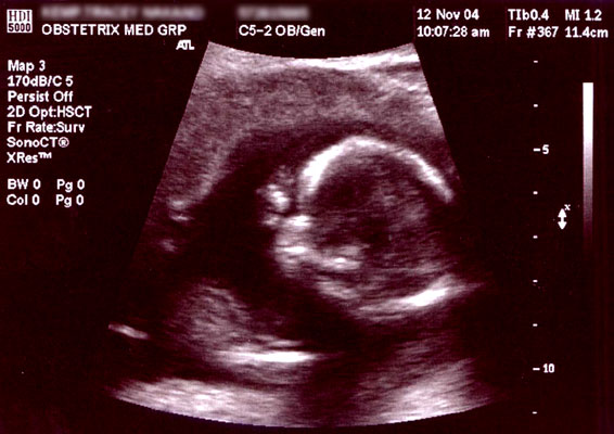
In my experience as an obstetrician, I have witnessed the immense impact that ultrasound technology has had on the field of obstetrics. Ultrasound has revolutionized prenatal care by allowing us to visualize the developing fetus in real-time and assess its well-being. This comprehensive guide aims to explore the various roles that ultrasound plays throughout pregnancy. From confirming the pregnancy in the early stages to monitoring fetal growth and development, ultrasound has become an indispensable tool in providing optimal care to expectant mothers. Additionally, ultrasound is instrumental in detecting any potential abnormalities or complications that may arise. By providing accurate and timely information, ultrasound empowers both healthcare professionals and parents to make informed decisions about the pregnancy. Overall, the advancements in ultrasound technology have undoubtedly transformed the way we approach obstetrics and have greatly improved the care we can provide to pregnant women.
2. Advancements in Ultrasound Technology: Revolutionizing Obstetric Imaging
As a woman, I have always been fascinated by the advancements in ultrasound technology and how it has revolutionized obstetric imaging. From the moment a woman finds out she is pregnant, ultrasound scans play a crucial role in monitoring the development of the fetus and ensuring a healthy pregnancy. With the advancements in ultrasound technology, doctors are now able to obtain more detailed images and even 3D and 4D scans, allowing parents to get a glimpse of their unborn child like never before. This not only strengthens the bond between parents and their baby but also aids in early detection of any abnormalities or complications. Additionally, ultrasound technology has made significant advancements in real-time imaging, allowing doctors to visualize the movements of the baby and assess its well-being. It is truly remarkable how far ultrasound technology has come and how it continues to improve the field of obstetric imaging.
3. Exploring the Benefits of Ultrasound in Cardiology: A Review
In my review of the benefits of ultrasound in cardiology, I have discovered how this imaging technique has made a significant impact in the field. Ultrasound allows doctors to visualize the heart and its structures in real-time, providing valuable information about its size, function, and blood flow. This non-invasive method has revolutionized the diagnosis and management of various cardiac conditions, such as heart valve disorders, congenital heart defects, and heart failure. Furthermore, ultrasound-guided procedures, such as echocardiography and Doppler imaging, have greatly improved the accuracy and safety of cardiac interventions, including catheterization and percutaneous coronary interventions. Overall, ultrasound has proven to be an invaluable tool in cardiology, enabling healthcare professionals like myself to better understand and treat cardiac diseases.
4. Ultrasound Imaging in Cardiology: An Essential Diagnostic Tool
Ultrasound imaging has revolutionized the field of cardiology, allowing doctors to obtain a clear and detailed view of the heart and its surrounding structures. As a cardiologist, I can attest to the essential role that ultrasound plays in the diagnosis and treatment of various cardiac conditions. By using high-frequency sound waves, this non-invasive procedure provides valuable information about the size, shape, and function of the heart, as well as the blood flow through its chambers and vessels. It allows us to identify any abnormalities, such as valve defects or blockages, and helps guide us in making informed treatment decisions. The real-time imaging capability of ultrasound also aids in the monitoring of patients during interventional procedures or surgeries. As the first female cardiologist in my department, I am proud to utilize this indispensable diagnostic tool to provide the best possible care to my patients.
5. How Ultrasound Helps in Detecting Fetal Abnormalities: Key Findings in Obstetrics
As an obstetrician, I am grateful for the advancements in ultrasound technology that have greatly improved our ability to detect fetal abnormalities. Ultrasound has become an indispensable tool in prenatal care, allowing us to gather valuable information about the developing baby’s growth and well-being. With the use of high-frequency sound waves, we can obtain detailed images of the fetus, enabling us to identify any potential abnormalities or developmental issues. This early detection is crucial as it allows for timely intervention and management, which can greatly improve outcomes for both the mother and the baby. Ultrasound has revolutionized the field of obstetrics, providing us with a non-invasive and accurate way to monitor the health of both the baby and the mother throughout pregnancy.
6. Innovations in Ultrasound Imaging: Improving Accuracy in Obstetrics and Cardiology
Innovation in ultrasound imaging has revolutionized the fields of obstetrics and cardiology, bringing about significant improvements in accuracy and diagnostic capabilities. As a physician specializing in obstetrics, I have witnessed firsthand the incredible advances in ultrasound technology. The introduction of 3D and 4D ultrasound has allowed us to visualize the fetus in incredible detail, providing invaluable information about its development and identifying any possible abnormalities. This has greatly enhanced prenatal care and the ability to intervene early if necessary. In cardiology, ultrasound imaging has become an essential tool for assessing the structure and function of the heart, aiding in the diagnosis and management of various cardiovascular conditions. These innovations in ultrasound imaging have undoubtedly transformed patient care in these crucial medical fields.
Conclusion
In conclusion, ultrasound imaging has revolutionized the fields of obstetrics and cardiology, providing valuable diagnostic tools that have greatly improved patient care. Obstetric ultrasound has allowed for the early detection of fetal abnormalities and provided valuable information for monitoring pregnancy. In cardiology, ultrasound has become an essential tool for assessing heart function and diagnosing various cardiac conditions. Overall, ultrasound imaging has proven to be an invaluable technology that continues to advance and benefit both patients and healthcare professionals in these specialized fields.
What is ultrasound imaging?
Ultrasound imaging, also known as sonography, is a medical imaging technique that uses high-frequency sound waves to produce visual images of the body’s internal structures.
How does ultrasound imaging work?
During an ultrasound exam, a transducer is used to send and receive sound waves. These waves bounce off the organs and tissues in the body, creating echoes that are captured and translated into images.
What are the benefits of ultrasound imaging?
Ultrasound imaging is non-invasive, painless, and does not use ionizing radiation. It can provide real-time images, allowing for immediate assessment. It is also useful for monitoring fetal development during pregnancy and diagnosing various medical conditions.
What are some common applications of ultrasound imaging?
Ultrasound imaging is widely used in obstetrics to monitor prenatal development, detect abnormalities, and guide procedures such as amniocentesis. It is also used in cardiology to assess heart function, detect cardiovascular diseases, and assist in procedures like echocardiography.
Are there any risks or side effects associated with ultrasound imaging?
Ultrasound imaging is considered safe and generally has no known risks or side effects. It does not use radiation like X-rays or CT scans. However, excessive use or prolonged exposure to ultrasound waves may cause tissue heating and potential harm to the fetus, so it should only be performed when medically necessary.
Is ultrasound imaging always accurate?
While ultrasound imaging is a valuable diagnostic tool, its accuracy may vary depending on factors such as the skill of the technician, the quality of the equipment, and the specific condition being evaluated. Sometimes, additional tests or imaging modalities may be needed to confirm a diagnosis.
